Glossary of Frequently Used Terms
| ablation | a minimally invasive technique that destroys varicose leg veins. The treatment sends bursts of radiofrequency or laser energy through a catheter; the resulting heat intentionally destroys vein wall tissues along the length of the vein. |
| angioma | a benign tumor made up of blood vessels or lymph vessels |
| angiomatosis | a condition characterized by multiple angiomas, or vascular tumors |
| anomaly | something that deviates from what is standard or expected |
| anticoagulant | a class of drugs that work to prevent the coagulation (clotting) of blood |
| arteriovenous fistula | abnormal connection between an artery and a vein |
| benign | not harmful, not malignant |
| bleb | small vesicles (fluid filled sac) that can appear, usually within the capillary lesion |
| capillary malformation | a flat, sharply defined vascular stain of the skin; malformed dilated blood vessels in the skin. Because the vessels are close to the surface, the increased flow gives the skin its pink to purple appearance. Reticular vascular naevi lighten over time, while port wine stains often deepen in color and the skin thickens. |
| cellulitis | bacterial skin infection that, left untreated, can potentially rapidly turn life-threatening. Cellulitis usually appears as a swollen, red area of skin that feels hot and tender. It can spread rapidly to other parts of the body. It is not usually spread from person to person. |
| chronic venous insufficiency | a condition that occurs when the venous wall and/or valves in the leg veins are not working effectively, making it difficult for blood to return to the heart from the legs. CVI causes blood to pool or collect in the veins, and this pooling is called venous stasis. |
| clotting factors | proteins in the blood that control bleeding. |
| collateral vein | the re-routing of blood circulation around a blocked artery or vein via nearby minor vessels |
| congenital disorder | a condition existing at or before birth regardless of cause |
| cutis marmorata | a condition where the skin has a pinkish blue mottled or marbled appearance when subjected to cold temperatures image |
| D-dimer | a fragment produced during the degradation of a clot. The D here stands for domain. Dimer indicates two identical units, in this case two identical domains. D-dimer result from complete breakdown of the clot. Monoclonal antibody to the D-dimer fragment provide the basis for the main methods of detecting it. The presence of D-dimers in the blood is a reliable clue that clotting has begun. Sometimes used as a clinical marker forpulmonary embolism (blood clot in the lung) or deep venous thrombosis (DVT) (blood clot in the leg) Sometimes written d-dimer or D-Dimer |
| differential diagnosis | the process of weighing the probability of one disease versus that of other diseases possibly accounting for a patient's illness. The differential diagnosis of rhinitis (a runny nose) includes allergic rhinitis (hayfever), the abuse of nasal decongestants and, of course, the common cold. |
| diffuse | not concentrated, spread over a large area; not limited or restricted |
| Deep Vein Thrombosis (DVT) | DVT occurs when a blood clot (thrombus) forms in one or more of the deep veins in your body, usually in your legs. Deep vein thrombosis can cause leg pain or swelling, but may occur without any symptoms. |
| embolization | a minimally invasive treatment that occludes, or blocks, one or more blood vessels or vascular channels of malformations (abnormalities). In a catheter embolization procedure, medications or synthetic materials called embolic agents are placed through a catheter into a blood vessel to prevent blood flow to the area. |
| embryogenesis | the development of a fertilized egg that occurs early on in pregnancy. After a sperm fuses with an egg, many changes occur in a specific order. The cells divide, reorganize and form layers of tissue that will eventually develop into specific organs. |
| embryonic vein | persistence of fetal veins beyond the normal atrophy during embryonic development |
| endometrial ablation | a procedure that surgically destroys (ablates) the lining of your uterus (endometrium). The goal of endometrial ablation is to reduce menstrual flow. In some women, menstrual flow may stop completely. The tools vary, depending on the method used to ablate the endometrium. They might include extreme cold, heated fluids, microwave energy or high-energy radiofrequencies. |
| endothelial cells | cells that line blood vessels |
| fibrin | a fibrous, non-globular protein involved in the clotting of blood. It is formed by the action of the protease thrombin on fibrinogen which causes it to polymerize. The polymerized fibrin together with platelets forms a hemostatic plug or clot over a wound site. |
| germline mutation | mutation (detectable variation) that occurs in the reproductive cells. Mutations in these cells are transmitted to offspring. |
| hemangioma | a benign and usually self-involuting tumor of the endothelial cells that line blood vessels characterized by increased number of normal or abnormal vessels filled with blood. Infantile hemangiomas usually appear in the first weeks of life and grows most rapidly over the first six months. Usually, growth is complete and involution has begun by twelve monthjs Half of all infantile hemangiomas have completed involution by age five, 70% by age seven, and most of the remainder by age twelve. Hemangiomas can interfere with vision, breathing, or threaten significant cosmetic injury. |
| hematuria | the presence of blood in the urine |
| hyperplasia | the enlargement of an organ or tissue caused by an increase in the reproduction rate of its cells, |
| hypertrophy | the enlargement of an organ or tissue caused by an enlargement of its component cells. Distinguished from hyperplasia, where cells remain the same size but increase in number. |
| hypotrophy | subnormal growth |
| interstitial | relating to, or situated in the small, narrow spaces between tissues or parts of an organ |
| interventional radiology | Interventional radiology (IR), also known as vascular and interventional radiology (VIR) or surgical radiology, is an independent medical specialty (previously a sub-specialty of radiology) providing minimally invasive image-guided diagnosis and treatment of diseases in every organ system. |
| lymphedema | an abnomal collection of high-protein fluid just beneath the skin that occurs when lymph vessels are damaged or nodes removed (secondary) or when lymphatic vessels are missing or impaired due to a congenital condition |
| malformation | abnormally formed part of the body |
| meninges | The meninges refer to the membranous coverings of the brain and spinal cord. There are three layers of meninges, known as the dura mater, arachnoid mater and pia mater. |
| mosaic/mosaicism | cells that undergo changes during development such that one group of cells differs from a neighboring group |
| Pulmonary embolism, PE | a blockage in one of the pulmonary arteries in your lungs. Most PEs are caused by blood clots (thrombosis) that travel to the lungs from another part of the body. |
| phlebectasia | the quality or state of being varicosed or dilated |
| phlebitis | see thrombophlebitis |
| phlebolith | small local, usually rounded, calcification within a vein. These are very common in the veins of the lower part of the pelvis, and they are generally of no clinical importance. |
| platelet | also called thrombocytes, platelets are tiny blood cells formed in bone marrow that help your body form clots to stop bleeding. If one of your blood vessels is damaged, it sends out signals that are picked up by the platelets, which then rush to the site of damage and form a plug, or clot, to repair the damage. |
| port wine stain | a type of capillary malformation also called a nevus flammeus. A large flat patch of purple or dark red skin with well defined borders. At birth the surface of the port wine stain is flat, but in time it becomes bumpy and takjes on a cobblestone appearance. They do not shrink or disappear. |
| primary lymphedema | a rare condition caused by problems with the development of lymph vessels in your body |
| prothrombin time (PT) | a blood test that measures how long it takes blood to clot. Can be used to check for bleeding problems or to determine if medicine to prevent blood clots is working. Also called INR test |
| reticulate vascular naevi | capillary malformations closely related to port wine stains. Reticulate vascular naevi often fade over time. |
| sclerotherapy | a treatment for small varicose and spider veins involving injecting a solution directly into the vein, causing the vein to scar and forcing blood to reroute through healthier veins. The collapsed vein is reabsorbed into local tissue and eventually fades. |
| scoliosis | sideways curvature of the spine |
| secondary lymphedema | lymphedema resulting from damage to lymph nodes or lymph vessels (surgery, radiation, infection) |
| somatic mutation | changes to the genetics not passed on to offspring through the germline |
| telangectasia | a condition characterized by dilation of the capillaries, which causes them to appear as small red or purple clusters, often spidery in appearance, on the skin or surface of an organ |
| thoracic duct | or the left lymphatic duct. The largest of the body's lymphatic ducts, it collects almost all the lymph circulating in the body. The rest is collected by the right thoracic duct. Lymph goes from there back into the bloodstream. |
| thrombocytopenia | the condition of having too few platelets, due to the bone marrow making too few platelets or the platelets being destroyed. If the platelet count gets too low, bleeding can occur under the skin as bruising, inside the body as internal bleeding, or outside the body through a cut that won't stop bleeding or from a nosebleed. Thrombocytopenia can be caused by many conditions. A normal platelet count is 150,000 to 450,000 platelets per microliter of blood. Risk for bleeding develops if a platelet count falls below 10,000 to 20,000. |
| thrombophlebitis, thrombosis | occurs when a blood clot blocks one or more vein. Thrombophlebitis (sometimes called phlebitis) can affect veins in the arms or neck. The affected vein may be near the surface of the skin, causing superficial thrombophlebitis, or deep within a muscle, causing deep vein thrombosis (DVT). |
| thrombus | blood clot |
| trophic changes | changes resulting from the interruption of nerve supplies |
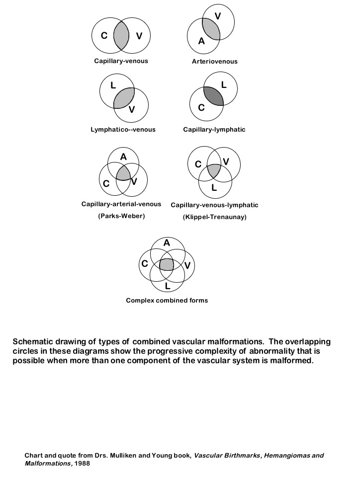
| vascular malformation | abnormal clusters of veins, lymph vessels, and/or arteries. CV, VM, LM, AVM or any combination (see Mulliken chart) |
| vein of Servelle (marginal vein of Servelle, lateral vein of Servelle | a persistent embryonic vein often apparent in Klippel Trenaunay syndrome. |
| venous stasis | slow blood flow, usually in the legs. Venous stasis is a risk factor for forming blood clots (thrombosis) or developing skin ulcerations. |
Page last updated April 8, 2016