Use this form to let us know if you are having difficulty with something on our website, or just to provide general feedback. This form will report your browser, device, and logged-in status to us along with your feedback.
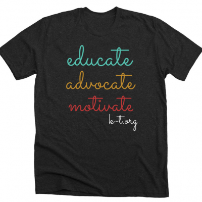
Support and resources for people with
Klippel-Trenaunay Syndrome
and related conditions.